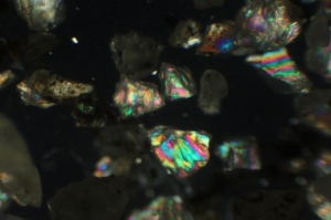
Hello fellow microscope hobbyists, I am starting my new post at blogger.com. See the pictures of my latest field trip mineral collection from Chucky Gal Mountain, North Carolina here.
Archive for the ‘Uncategorized’ Category
The blog is moving
Posted in Microscope, Projects, Uncategorized on June 6, 2011| Leave a Comment »
Amos Cunningham Farm Field Trip
Posted in Mineral and Rocks, Uncategorized on October 2, 2010| Leave a Comment »
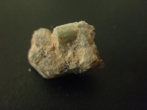
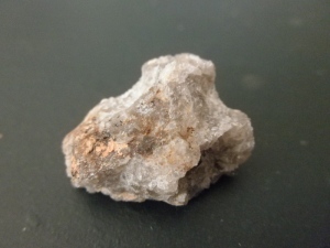
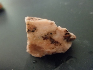
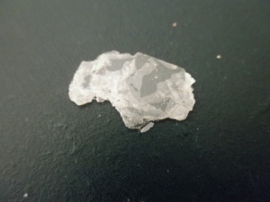
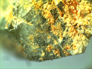
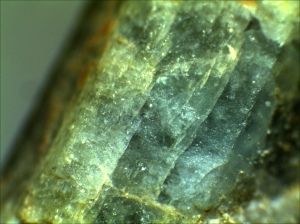
The destination of my second Georgia Mineral Society field trip is Amos cunningham Farm located in Due West/Antreville Area, South Carolina. The collection site is known for its beryl, amethyst and smoky quartz. The site is flat to rolling land of South Carolina red clay. The digging areas have been machine trenched down to white kaolite veins that have the beryl and quartz crystals. Dirt and rock removed from the trenches and piled up, also contain beryl and are good places to search.
Some macro shot of the specimen collected in the field trip:
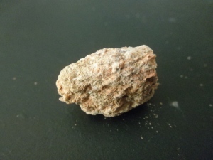
Aside from felspar, there are also plenty of Pumice in the kaolite vein. Pumice is a very porus and light rock. If you put them in water, you can heal the bubbling sound.
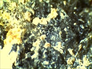
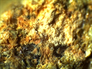
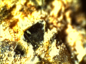
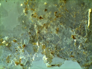
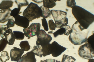
Images of the collection under stereo microscope:
The macro shot was captured by Olympus Stylus 7010 camera in super macro mode. The photomicrograph was the specimens are taken with Tucsen Microscope Camera from Ample Scientific SM-Plus-24 Stereo Microscope.
Collectings Insects with Lights
Posted in Uncategorized on July 7, 2010| Leave a Comment »
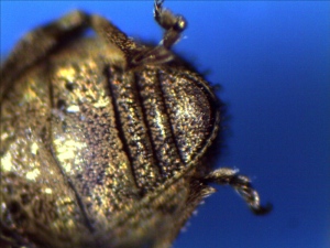
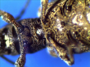
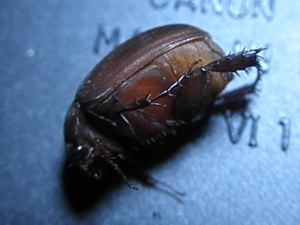
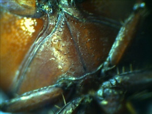
Insects are attracted to light to navigate. This behavior have been evolved million years ago when the only light during the night are moon. Insects will fly directly toward the moon. This instinct causes insects to fly toward artificial lights. Insects are particularly attracted to light with shorter wave lengh, such as black light – You can obtain the black light tubes from pet store which they are sold to detect pet urines around the house. If you don’t have black light, you can use regular fluorescent light or halogen lights or simply trolling under the street lights. You will be surprised how many bugs you can find.
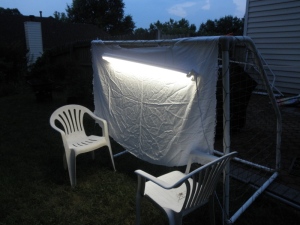
Collecting Insects with Light. If you don't have black, a regular fluorescent will do the job. I put a white cloth to reflect the light. It will also allow you easier to spot the visitors.

An insect collection tube is helpful to keep the small insects. You might also bring a killing jar for larger insects.
What did I found?

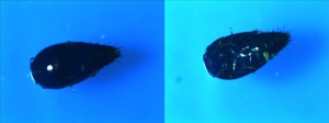
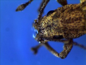
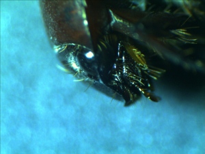
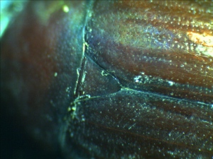
Rove bettle is characterized by very short elytra and leave more then half or their abdomen exposed.
Photos are taken with Tuscen 3.0 MP CMOS microscope camera through the eyepiece of Ample Scientific SM Plus Stereo Microscope. The amplificion is either 20 x or 40x
Related Posts:
My first attempt of making insect slides (of ants and aphids) using common household chemicals at my new practical microscopy blog at http://practical-microscopy.blogspot.com
Using Tucsen Microscope Camera with Carl Zeiss AxioVision LE
Posted in Microscope Camera, Uncategorized, tagged AxioVision, Image Acquisition Software, MICAM, Microscope Camera, TSview, Tucsen on June 19, 2010| 1 Comment »
Carl Zeiss AxioVision is the image acquisition and processing software developed by optical equipment giant, Carl Zeiss Company. This software is a very comprehensive and powerful software. The commercial version is commonly bundled with Zeiss AxioCam and microscopes. The retail price for the software is $1,200 but there is a free downloadable feature limited version, AxioVision LE. The current version is 4.8.1. You can download it from Zeiss website, http://www.zeiss.de/axiovision.
The installation is very straight forward just like any othe windows applications (I first request a download link from Zeiss AxioVision website then run the executable from the downloaded file -about 230mb). The installation wizard took me through the installation process. It took less than 10 minutes. To use the Zeiss AxioVision with your Tucsen microscope camera, you want to enable the DirectShow component from the Module Manager. To get to the the module manager, select Tools -> Module Manager from the main menu.
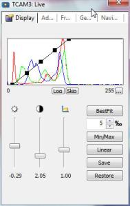
The interface of the AxioVision is very user friendly. The layout of the application stayed with windows application convention so it will give you a familar feeling with other application. There isn’t much hidden funtion which you can’t get to it in a couple mouse click. The main menu and toolbar is across the top. The explore view on the left allow you to select different options (such as camera, image capturing and processing). After you click on the option, you can configure the options on the bottom. Click on the Live on the toolbar will allow you to see the preview window and click on the Snap will allow you to take a picture. In the live preview window, you can configure various image acquisition options. The best fit is my favorite option which allow you to stretch the colors to full dynamic range. The image is vibrant and have live in it. The histogram is very useful. It will take away the guessing work out when you have to do manual adjustment of image acquisition.
How does AxioVision LE perform?
Under low light condition
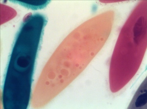
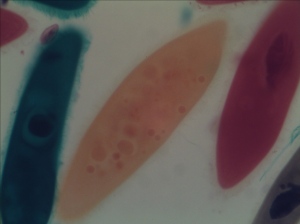
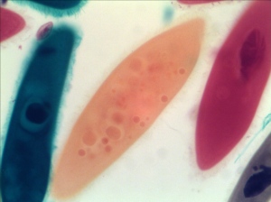
Some After Thoughts The images acquired by AxioVision is very colorful and very close to actual view (except a little too much brown/orange on the paramecium stained with yellow. The red and green color are both very truthful. It seems to be able to adjust the brightness. There isn’t much difference between low light and normal light condition. The best fit is what makes it stand out compared to TSView and MICAM. This is the type of post-processing that I will consider to be added to the image captured by either TSView or MICAM. Now you can do it with AxioVision LE in one snapshot!
The Axiovision comes with many configuration options beyond the simple image acquisition. It is the software that I will be highly recommend the Tuscen users to try it out. The only downside? There isn’t any video capturing option so you can only use it to take snapshot pictures.
Damselfly
Posted in Pond Life, Uncategorized, tagged Damselfly under microscope, Nexcope microscope, Pond Life, Tucsen on June 11, 2010| Leave a Comment »
Damselfly is an aquatic insect. Their naiads live in water. They develop through 10 to 12 immature stages (instars), depending on the species and habitat. The last immature stage crawls out of the water onto vegetation before the adult emerges. The adults emerge from water and live for a few weeks to a few months. Mating is unusual: males deposit sperm in a secondary genitalia structure on the second and third abdominal segment by bending the abdomen forward. The male then clasps the female behind the head with claspers on the tip of his abdomen and mating pairs can be seen flying in tandem. The female then loops her abdomen forward and picks up the sperm from the male. Eggs are deposited in emergent plants or floating vegetation or directly into the water.
Nymph picture:

damselfly naiad - picture was taken at 40X through the eyepiece of Nexcope CM701 microscope with Tucsen microscope camera
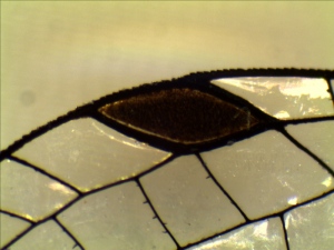
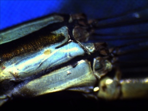
Adult damselfly (Turquois Bluet damselfly – Enallagma divagans) under microscope
This damselfly appears to be a female. There are two spikes on the anal appendges but it seems to be too short to hold the female for mating. The second segment of the abdomen does not have the structure for dispensing the sperms.
Damselfly are closely related to anothe aquatic insects, dragonfly. The damselfly has:
- long and slender body
- eyes are clearly separated
- all wings are in similar shape and size
- when rest the wings are held close and upright on the top of the abdomen
- The naiad are also slender and breath through gills
The dargonfly:
- usually stocky
- eyes are touched and on the top of the head
- Dissimilar wing pairs
- when rest, the wing held open, horizontally or downward
- The naiad has stock body and breath through rectal tracheal gill
You are are living in Georgia, USA or southeast georgia. Here are the website for our local dragonfly and damselfly websites:
Dragonfly and Damselfly in Georgia
Dragonfly and Damselfly of Georgia and southeast by Giff Beaton



















 Free Living FreshWater Protozoa by DJ Patterson
 How to Know the Protozoa
 Ecology and Classification of North American Freshwater Invertebrates by Thorp and Covich