Feeds:
Posts
Comments

Posts Tagged ‘Microscope’

The subjects of my interests about microscopy changes from time to time.  I have been crazy about rocks and minerals this summer.   The temperature here has been dipped down to mid 40s degree F (7 degree C) in the early morning although the mid day temperature is still comfortably at mid 70s.  I got to go out to collect some pond specimens for observation before it is too cold for any organism to be active.  I choose different collection site, Briscoe Park Located at Snellville, GA.

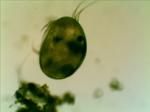
The number and variety have been drastically reduced from my early summer observation.  Again, I found seed shrimp (Ostracod) in my collection.

Seed Shrimp (Ostracod)

Seed Shrimp (Ostracod)

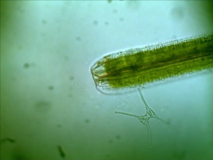
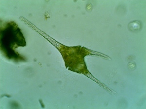
A Dinoflagellate Ceratium spp.

First time, I observed a dinoflagellate (Ceratium Furca). It is a flagellate protist. I can barely see the flagella because the flagella are relatively transparent.

Diatom

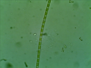
filamentous Green Algae

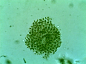
Unidentified Algae Colony

The specimen was collected in 60 mL tube and split into a several 7 mL tubes and centrifuged in my homemade centrifuge.

Read Full Post »

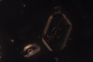
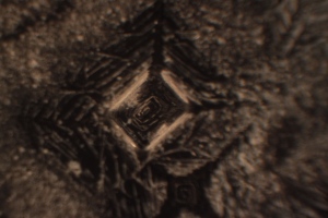
dissolve mineral and crystalized the minerals is another way to prepare the specimen for polarized light microscope examination.  Yesterday, I dissolve several common ingredients for food seasonings and put a few drops on the slides to allow them to dry: table salt (Sodium Chloride), Sugar and Alum hydrated potassium aluminium sulfate.  I also drop a few drops of methylene blue.  Today they are all pretty much dried out – The sugar is still somewhat sticky but I can clearly see the sugar crystals in snow flake shape.

White Sugar Crystal under Polarized Light Microscope

White Sugar Crystal under Polarized Light Microscope

White Sugar Crystal under Polarized Light

White Sugar Crystal under Polarized Light (This is the first photo with some post processing by adjusting the gamma to 0.6 from 1.0)

 The color of the sugar crystal shows richer color when I adjust the gamma correction.  The loss of color must have been caused by the manual shooting mode. 

White sugar crystal under polarized light microscope

White sugar crystal under polarized light microscope

White sugar crystal under polarized light microscope

White sugar crystal under polarized light microscope

Alum crystal under polarized light microscope

Alum crystal under polarized light microscope

Table salt under polarized light microscope

Table salt under polarized light microscope

Methylene blue under polarized light microscope

Methylene blue under polarized light microscope

The photos are taken with Canon EOS T1i with Manual mode (ISO 400, exposure 4 sec, auto white balance).  Nexcope CM701 converted polarized light microscope was used for observation.

Read Full Post »

Guppies (Poecilia reticulata)  is the most popular aquarium fish.  They are community fish which means that they get alone with other aquarium fish.  They are not very picky about water condition and easy to breed.  I have a tank of guppies that I have kept for several generations.  

Guppies (Poecilia reticulata) are peaceful community fish

Guppies (Poecilia reticulata) are peaceful community fish

Fish fins are transparent or semi-transparent.  They can be easily observed under microscope without any sample preparation. I took five guppies from my fish tank and put them in a small container next to my microscope so I can easily put the fish back in the water (They can’t be leave out of water for extended period of time).  I lay the fish on a slide then put a piece of moistened cotton ball on the head.  It servers two purpose: 1. Put some weight on the fish so it wouldn’t jump. 2. Prevent it from drying out too quickly (The fish won’t be able to breathe but I think a little bit moisture will help).  I tried to put the cover slide on but they were quickly move by fish’s powerful tail.  I end up not using the cover slide.  For this reason, the observation is only limited to 40X and 100X.  The working distance of 40X objective is too short. 

Guppy's fin at 40X

Guppy's fin at 40X

Guppy's fin at 100X

Guppy's fin at 100X

The blood circulation is recorded at 100X with Tucsen Microscope Camera.

Read Full Post »

I left a piece of rotten peach indoor.   A couple days later, I noticed very dense of mycelia were growing on it.  I put it under the microscope, there are may spore producing structure on the tip of the stalk.

Monila fructicola at 40X under stereo microscope. The black dots are the sporangia of Monilinia fructicola.

Sporangia of Monilinia fructicola at 400x. The specimen is stained with Malachite Green.

A sporangium of Monilinia fruticola at 400x. (The photo was cropped to show better details.)

The original post of the Peach Brown Rot Monilinia fruticola

Read Full Post »

Brown Rot is a serious disease for peach trees.  This disease is caused by fungus, Monilinia fructicola.  The M. fructicola can infect flowers, shoots, fruits and branches.  Typical disease symptoms induced by M. fructicola include blossom and twig blight, cankers, and a fruit rot.   Brown rot on the peach typically develops rapid brown necrosis.  Under favored condition, the entire fruits can be rotten within 48 hours of infection. 

M. fructicola over winters in dried infected fruit called peach mummies or in infected branch cankers.  Mummies can either remain on tree or scatterd on ground.  Both may produce spores which infect blossoms and young fruit in the spring.  There are wo types of spore: ascospores and conidia. Ascospores are produced from apothecia, a mushroom-like structure that occurs only on mummies which have fallen to the ground and are partially covered with soil. Conidia are produced in abundance on mummies and infected twigs and may be spread by wind and rain. 

Here at Georgia, nicknamed peach state, brown rot is the serious disease for commercial peach orchard.  The peach tree in my backyard is not immuned from the disease.  There are beautiful peach blossom during the spring but what follows is the ugly brown rot and fruit droppings all over the ground. 
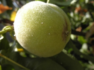
Peach insect bites

The symptoms on the fruits seems to start from a small insect bite where a small brown dot and clear gel like dischrage is clearly visible.

Brown Rot and Fruit Drop

The brown rot was spread to half of the fruit. There are fruits droppings every where on the ground.

peach mummy

A completely rotten peach is hanging on the tree. It is also called peach mummy.

Another peach mummy

I collected some peach infected by brown rot and slice a thin layer of skin.  Put it on my newly acquired Ample Scientific SM Plus stereo microscope with only the top light on. 

Healthy Peach Skin aT 20x

Healthy Peach Skin at 20x.

The skin of brown rot infested peach. The skin turned brown even without clearly visible mycelium.

Peach pulp and skin

Peach pulp and skin

Peach Pulp

The pulp of a peach infected by brown rot. Half of the pulp has already turned brown.

Monilinia fructicola Mycelium

The mycelium of Monilinia fructicola on a peach mummy.

A piece of peach mummy skin was sliced off and placed it in a vial with small amount of water. I gently scrap the surface with a dissecting tweezer, capped the vial and shacked for a few seconds.  A simple wet mount and malachite green stain was applied to the specimen.  The slide then observed at 400X with Nexcope CM701 compound microscope. 

Conidia of Monilinia fructicola

The pathogen that causes the peach brown rot. The picture is the conidia of Monilinia fructicola

two conidiospores formed on conidia (Monilinia fructicola)

Conidia of Monilinia fructicola

The specimens of conidia of Monilinia fructicola were stained with Malachite green.  Photos were taken with Tucsen 3.0MP CMOS microscope camera using Zeiss AxioVision 4.8 LE image capture software.  The eyepiece of the microscope was removed and replaced with Tucsen microscope camera adapter. 

Couple days after I left the rotten peach in door.  Rotten peach continued…

More web links about peach brown rot: 

University of Georgia Peach Handbook – Brown Rot

Cornell University Tree Fruit and Berry Pathology 

University of West Virginia KTFREC – Plant Disease Fact Sheet

Read Full Post »
Dragonflies belong to the Odonata order.  It is characterized by very large mutifaceted eyes, two pair of strong transparent wings and elongated wings.  Dragonflies are similar to damselflies, but the adults can be differentiated by the position of their wings while resting.  Dragonflies’ wings are held to the side and perpendicular to the body while the damselflies held their wings in verticle position.

Dragonflies usually has a life span of more than a year.  The adult dragonflies mate in the air.   After them mate, the female dragonflies lay eggs on the plant in the water.  If they can’t find a suitable plant, they will just drop them in water.  The nymph stage begin after the egg hatches. Dragonfly lives in the water while they grow and developed into dragonfly.   The nymph stage can take up to four years to complete.  The dragonfly nymph will come out of water and turn into adult, once the dragonfly nymph fully grown and the temperator is warm.   They will hunt and start to mate.  The adult stage typically lasts less then two month.

You should not have trouble finding in near by pond during summer since dragonflies are aquatic insects.  We collected a Eastern Amber Wing dragonfly (Perithemis tenera) near a pond at little mulberry park located in Dacula, GA, USA on Jun 26, 2010.

Photos of dragon under microscope

Dragonfly

Dragonfly (the end of the abdomen was chewed off by a Katydid whiling fighting in the collecting jar)

Mouth part of a dragonfly

Mouth part of a dragonfly (This is a stacked photo from 3 photos)

Dragonfly's head

Dragonfly's head

The front edge of the wing of a dragonfly

The front edge of the wing of a dragonfly

Wing of dragonfly

Wing of dragonfly

Front leg and compound of a dragonfly

Front leg and compound of a dragonfly

Photos are taken with Tuscen 3.0 MP CMOS microscope camera through the eyepiece of Ample Scientific SM Plus Stereo Microscope.  The amplificion is either 20 x or 40x.

Related post:

My first attempt of making insect slides with common household chemicals at my new blog at http://practical-microscopy.blogspot.com.  The pictrue below is an aphid’s head and thorax. 

The head and thorax of an aphid

The head and thorax of an aphid

Read Full Post »

Older Posts »